Question:
A child presented to the hospital with cola-colored urine, hypertension, and puffiness of eyes. Laboratory investigations were done, and creatinine was 2.5 mg/dL. Treatment was started, and despite treatment, the patient did not improve for the next 3 weeks. The creatinine value increased to 4.5 mg/dL. Which among the following electron microscopic findings will be seen in this patient ?
A child presented to the hospital with cola-colored urine, hypertension, and puffiness of eyes. Laboratory investigations were done, and creatinine was 2.5 mg/dL. Treatment was started, and despite treatment, the patient did not improve for the next 3 weeks. The creatinine value increased to 4.5 mg/dL. Which among the following electron microscopic findings will be seen in this patient ?
Updated On: Jun 19, 2025
Subendothelial deposits
- Subepithelial deposits
Crescent formation
Mesangial deposits
Hide Solution
Verified By Collegedunia
The Correct Option is B
Solution and Explanation
The clinical presentation described in the question suggests the possibility of acute post-streptococcal glomerulonephritis (APSGN), particularly given the cola-colored urine (indicative of gross hematuria), hypertension, and edema (puffiness of eyes). APSGN typically occurs following an infection with group A beta-hemolytic streptococcus. Regarding the laboratory tests, the elevated creatinine levels (from 2.5 mg/dL to 4.5 mg/dL) indicate worsening renal function, which aligns with significant glomerular injury.
To determine the electron microscopic findings, we need to understand the characteristics of APSGN:
- APSGN characteristics: It is an immune complex-mediated disease where antigen-antibody complexes deposit in the glomeruli. Classically, APSGN is associated with subepithelial immune complex deposits, known as "humps," which can be identified using electron microscopy.
| Characteristic Finding in APSGN | Subepithelial deposits |
|---|
Thus, given this clinical scenario and the understanding of APSGN, the electron microscopic finding most likely seen in this patient is subepithelial deposits, aligning with the correct answer from the options provided.
Was this answer helpful?
0
0
Top Questions on Pediatric Disorders
- A 48-year-old man presents with complaints of facial puffiness, frothy urine, and hypertension. He gives a history of infection with hepatitis B. Urine examination reveals microscopic hematuria. The histopathological image of the kidney biopsy shows a spike and dome pattern. What is the diagnosis of this condition ?
- NEET (PG) - 2023
- Pathology
- Pediatric Disorders
- A 3-month-old baby presents with jaundice and clay-coloured stools. Lab investigation reveals that the baby has conjugated hyperbilirubinemia. The liver biopsy shows periductal proliferation. What is the most likely diagnosis?
- NEET (PG) - 2023
- Pediatrics
- Pediatric Disorders
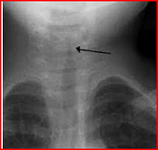
- A child is brought to the hospital with respiratory distress and biphasic stridor. The radiograph is shown below. What is the diagnosis?
- NEET (PG) - 2023
- Pediatrics
- Pediatric Disorders
- An infant is brought with complaints of excessive watering of the eyes and photophobia. The image is given below. What is the likely diagnosis ?

- NEET (PG) - 2023
- Ophthalmology
- Pediatric Disorders
Chloride level in sweat is used in the diagnosis of which disease?
- NEET (PG) - 2023
- Pediatrics
- Pediatric Disorders
View More Questions
Questions Asked in NEET PG exam
Which of the following cranial nerves is responsible for the motor innervation of the muscles of mastication?
- NEET (PG) - 2025
- General Science
The normal pH of arterial blood is:
- NEET (PG) - 2025
- General Science
Which enzyme is deficient in Gaucher’s disease?
- NEET (PG) - 2025
- General Science
The anticoagulant effect of heparin is monitored using:
- NEET (PG) - 2025
- General Science
The causative agent of malaria is:
- NEET (PG) - 2025
- General Science
View More Questions