Question:
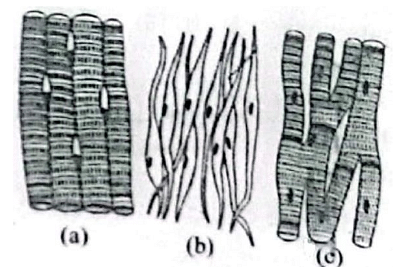
Three types of muscles are given as a, b and c. Identify the correct matching pair along with their location in human body :

Three types of muscles are given as a, b and c. Identify the correct matching pair along with their location in human body :


Updated On: Jan 13, 2026
- (a) Smooth - Toes
(b) Skeletal – Legs
(c) Cardiac – Heart - (a) Skeletal - Triceps
(b) Smooth – Stomach
(c) Cardiac – Heart - (a) Skeletal - Biceps
(b) Involuntary – Intestine
(c) Smooth – Heart - (a) Involuntary – Nose tip
(b) Skeletal – Bone
(c) Cardiac – Heart
Hide Solution
Verified By Collegedunia
The Correct Option is B
Approach Solution - 1
In the context of human biology, the three main types of muscle tissues are skeletal, smooth, and cardiac, each having distinct characteristics and locations in the body.
- Skeletal Muscles: These are voluntary muscles attached to bones and are responsible for body movement. A common example is the muscles in the arms, such as the triceps and biceps.
- Smooth Muscles: These are involuntary muscles found in the walls of internal organs like the stomach, intestines, and blood vessels. They help in various functions like digestion and regulation of blood flow.
- Cardiac Muscles: These specialized muscles are found only in the heart. They are responsible for the continuous contraction necessary to pump blood throughout the body.
Given the options, the correct matching pair, based on the characteristics and location of each muscle type, is:
| (a) Skeletal - Triceps |
| (b) Smooth – Stomach |
| (c) Cardiac – Heart |
Was this answer helpful?
4
2
Hide Solution
Verified By Collegedunia
Approach Solution -2
Classification of Muscle Types:
Skeletal muscle – Found in the triceps and is voluntary in nature. (a)
Smooth muscle – Found in the walls of the stomach and is involuntary. (b)
Cardiac muscle – Found in the heart and is involuntary. (c)
Thus, the correct answer is (2): (a) Skeletal – Triceps, (b) Smooth – Stomach, (c) Cardiac – Heart.
Was this answer helpful?
0
0
Top Questions on Human body
- Which of the following is not a part of the human digestive system?
- CUET (UG) - 2025
- Biology
- Human body
- The part of the brain responsible for maintaining posture and balance is the:
- CUET (UG) - 2025
- Biology
- Human body
- Which part of the nephron is primarily responsible for filtration of blood?
- CUET (UG) - 2025
- Biology
- Human body
- The large bean-shaped organ acting as a filter of the blood in humans is:
- Given below are two statements:
Statement I: The cerebral hemispheres are connected by nerve tract known as corpus callosum.
Statement II: The brain stem consists of the medulla oblongata, pons and cerebrum.
In the light of the above statements, choose the most appropriate answer from the options given below:- NEET (UG) - 2024
- Biology
- Human body
View More Questions
Questions Asked in NEET exam
- Two cities X and Y are connected by a regular bus service with a bus leaving in either direction every T min. A girl is driving scooty with a speed of 60 km/h in the direction X to Y. She notices that a bus goes past her every 30 minutes in the direction of her motion, and every 10 minutes in the opposite direction. Choose the correct option for the period T of the bus service and the speed (assumed constant) of the buses.
- NEET (UG) - 2025
- Relative Velocity
- A physical quantity P is related to four observations a, b, c, and d as follows: P = a3 b2 (c / √d) The percentage errors of measurement in a, b, c, and d are 1%, 3%, 2%, and 4% respectively. The percentage error in the quantity P is:
- NEET (UG) - 2025
- Dimensional analysis and its applications
What is Microalbuminuria ?
- NEET (UG) - 2025
- Human physiology
The output (Y) of the given logic implementation is similar to the output of an/a …………. gate.

- NEET (UG) - 2025
- Logic gates
- An oxygen cylinder of volume 30 litre has 18.20 moles of oxygen. After some oxygen is withdrawn from the cylinder, its gauge pressure drops to 11 atmospheric pressure at temperature \(27^\circ\)C. The mass of the oxygen withdrawn from the cylinder is nearly equal to: [Given, \(R = \frac{100}{12} \text{ J mol}^{-1} \text{K}^{-1}\), and molecular mass of \(O_2 = 32 \text{ g/mol}\), 1 atm pressure = \(1.01 \times 10^5 \text{ N/m}^2\)]
- NEET (UG) - 2025
- Ideal-gas equation and absolute temperature
View More Questions