Question:
A patient presents to the clinic with the lesion given in the image. He had a traumatic injury to the chest one year ago. What is the most likely diagnosis?
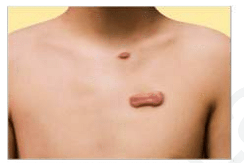
A patient presents to the clinic with the lesion given in the image. He had a traumatic injury to the chest one year ago. What is the most likely diagnosis?
Updated On: Jun 18, 2025
- Hemangioma
- Hypertrophic scar
- Keloid
- neurofibroma
Hide Solution
Verified By Collegedunia
The Correct Option is C
Solution and Explanation
The case presents with a lesion following a traumatic chest injury one year ago. To identify the most likely diagnosis, let us understand the characteristics of each condition:
- Hemangioma: Typically presents at birth or shortly thereafter, not usually associated with trauma.
- Hypertrophic Scar: Raised, red scars that remain within the boundaries of the original wound, often developing shortly after injury.
- Keloid: Raised, thick, irregular scar tissue that extends beyond the original wound boundary. Often develops months to years after injury.
- Neurofibroma: Usually presents as soft, fleshy lesions associated with nerves, not commonly linked to trauma.
Given the history of a traumatic injury and the development of a lesion that may exceed the wound's original boundaries, the most likely diagnosis is a Keloid. Unlike hypertrophic scars, keloids are known for growing beyond the original site of skin damage, often occurring long after the inciting trauma.
| Condition | Characteristics |
|---|---|
| Hemangioma | Common in infancy |
| Hypertrophic Scar | Confined, early formation |
| Keloid | Extends beyond wound, late formation |
| Neurofibroma | Associated with nerve fibers |
Therefore, given the patient's history and lesion behavior, the likely diagnosis is Keloid.
Was this answer helpful?
1
0
Top Questions on Plastic Surgery
- What is the ideal pressure setting for vacuum-assisted closure (VAC) therapy?
- NEET (PG) - 2024
- Surgery
- Plastic Surgery
- Sushrut first performed plastic surgery on:
- BCECE Nursing - 2024
- Biology
- Plastic Surgery
- Identify the given condition
- NEET (PG) - 2023
- Surgery
- Plastic Surgery
- Which of the following is false about the given condition?
- NEET (PG) - 2023
- Surgery
- Plastic Surgery
Questions Asked in NEET PG exam
Which of the following cranial nerves is responsible for the motor innervation of the muscles of mastication?
- NEET (PG) - 2025
- General Science
The normal pH of arterial blood is:
- NEET (PG) - 2025
- General Science
Which enzyme is deficient in Gaucher’s disease?
- NEET (PG) - 2025
- General Science
The anticoagulant effect of heparin is monitored using:
- NEET (PG) - 2025
- General Science
The causative agent of malaria is:
- NEET (PG) - 2025
- General Science
View More Questions