Question:
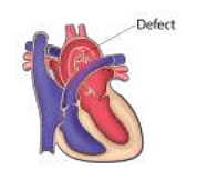
A defect in which of the following aortic arches causes the defect shown in the image?
A defect in which of the following aortic arches causes the defect shown in the image?
Updated On: Jun 18, 2025
4th right
4th left
6th right
6th left
Hide Solution
Verified By Collegedunia
The Correct Option is D
Solution and Explanation
The image reflects a defect associated with the development of the aortic arches in embryology. To determine the specific arch responsible for the defect, it's essential to understand the anatomy and the derivatives of these arches. During human development, several aortic arches form and then remodel into different structures:
- The 4th right aortic arch contributes to the right subclavian artery.
- The 4th left aortic arch contributes to the aortic arch (between the left common carotid and the left subclavian arteries).
- The 6th right aortic arch contributes to the right pulmonary artery.
- The 6th left aortic arch contributes to both the left pulmonary artery and the ductus arteriosus.
Was this answer helpful?
0
0
Top Questions on Embryology
- Select the mismatched pair: a) First month of pregnancy-Formation of heart
b) Second month of pregnancy-Movement of foetus
c) Third month of pregnancy-Formation of most of the major organ systems
d) Sixth month of pregnancy-Eye lids separate and eye lashes are formed- KCET - 2025
- Biology
- Embryology
Read the following statements: Statement I: All vertebrates develop a row of vestigial gill slits during embryonic stage.
Statement II: Embryos always pass through the adult stages of other animals.- KCET - 2024
- Biology
- Embryology
- The covering of an omphalocele is derived from which layer?
- NEET (PG) - 2024
- Anatomy
- Embryology
Which of the following is true regarding the image provided?

- NEET (PG) - 2024
- Anatomy
- Embryology
- The cavity of blastula is
- UPCATET - 2024
- Biology
- Embryology
View More Questions
Questions Asked in NEET PG exam
Which of the following cranial nerves is responsible for the motor innervation of the muscles of mastication?
- NEET (PG) - 2025
- General Science
The normal pH of arterial blood is:
- NEET (PG) - 2025
- General Science
Which enzyme is deficient in Gaucher’s disease?
- NEET (PG) - 2025
- General Science
The anticoagulant effect of heparin is monitored using:
- NEET (PG) - 2025
- General Science
The causative agent of malaria is:
- NEET (PG) - 2025
- General Science
View More Questions